epidural didn t work
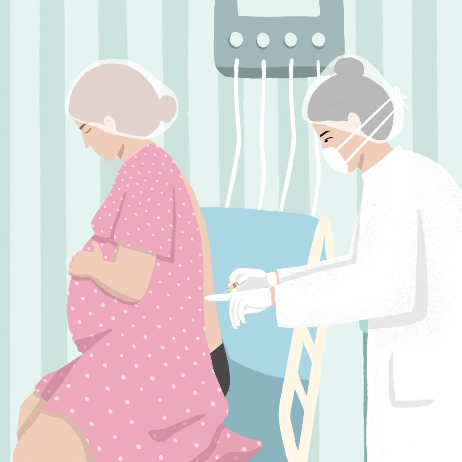 My Epidural Didn't Work. Here's What I Wish I Knew | Parents
Warning: The NCBI website requires JavaScript to operate. Why do epidurals not always workKatherine Arendt*Department of Anesthesiology, Mayo Clinic, Rochester, MNScott Segal†Department of Anesthesiology, Perioperative Medicine and Pain, Brigham and Women's Hospital, Harvard Medical School, Boston, MAAbstract The overwhelming majority of epidural catheters placed for work provide a satisfactory analgesia. However, there are times when the catheter is not properly settled within the epidural space, the patient's neuroraxial anatomy is problematic, or the patient's work progresses faster than expected by the anesthesiologist, and the epidural block is not established on time. In this article, the foundations of the pneumaxal work analgesia, the causes of their failure and the strategies that anesthesiologists use to rescue malfunction catheters are reviewed. A 28-year-old primitive term presents painful work. A 3 cm cervical dilation is placed after multiple attempts by an experienced anesthesiologist. Upon returning 2 hours later for a cervical examination, the obstetrician observes a parturant in the extremis, blinking and groaning in pain and denying the vaginal examination. The anesthesiologist is notified. Fortunately, the overwhelming majority of epidural catheters placed for work provide satisfactory analgesia. In large series, the overall success rate is approximately 98% to 99%. However, there are times when the catheter is not properly settled within the epidural space, the patient's neuro-axial anatomy is problematic, or the patient's work progresses faster than expected by the anesthesiologist, and the epidural block is not established on time. In this article we review the foundations of the pneumorial work analgesia, the causes of their failures, and the strategies that anesthesiologists use to rescue malfunction catheters. Neutral Anatomy and Epidural Technique The epidural space is a potential space in the back of neuraxis (previous) to the flavum ligamentum and superficial (after) to the hard mat. Anesthesiologists use a technique of loss of resistance to determine when the tip of a hollow epidural needle was located just before the thick ligament flavum and in the epidural space (). When the tip of the epidural needle is found within a ligament, the injection of air or saline is difficult. When entering the epidural space, this resistance to injection is lost. An epidural catheter is then threaded from 3 to 5 cm in the epidural space (). After the catheter sits, epidural drugs are injected and slowly absorbed into the nerve roots of the lower thoracic, lumbar and sacral column, providing analgesia in about 20 minutes. Epidural needle in the epidural space. De Eltzschig HK, Lieberman ES, Camann WR. Regional anesthesia and analgesia for childbirth and childbirth. N Engl J Med. 2003;348:319–332. Copyright © Massachusetts Medical Society. All rights reserved. Catheter placement in epidural space. De Eltzschig HK, Lieberman ES, Camann WR. Regional anesthesia and analgesia for childbirth and childbirth. N Engl J Med. 2003;348:319–332. Copyright © 2003 Massachusetts Medical Society. All rights reserved. In the epidural technique (CSE), after the epidural needle is well sida, a small, increased spinal needle is passed through the hole of the epidural needle, through the hard matt, and in the intratecal space (). Brain spinal fluid (CSF) flow through the spinal needle confirms its location. After the medication is deposited in the CSF, the spinal needle is removed, and an epidural catheter is threaded through the epidural needle in the epidural space. Intratecal medicine provides a faster appearance of analgesia than a single epidural, usually in 5 minutes. Combined spinal epidural technique. De Eltzschig HK, Lieberman ES, Camann WR. Regional anesthesia and analgesia for childbirth and childbirth. N Engl J Med. 2003;348:319–332. Copyright © 2003 Massachusetts Medical Society. All rights reserved. Neurological drugsBoth opioids and local anesthetics are typically used in the neuronal work analgesia. Phentanile is usually the opioids of choice because its lipofilicity results in an excellent immediate agonism of the muopioid receptor in the neurox and minimal central side effects. Bupivacaine is the local anesthesia of choice because, compared to other local anesthesias, it preferentially blocks motorized nerves, preserving the lower extremity and abdominal muscle strength. The concentration of volume and drugs should be considered in epidural dosing, while the baricity of the drug (density compared to CSF) within the CSF and the general dose (milligrams of drugs) should be considered in the intratecal space. The required dose in the epidural versus intratecal space differs approximately in a factor of 10. An epidural catheter placed without knowing it in the intratecal space could lead to massive overdose, which could lead to a "high bone". When this occurs, respiratory problems, hypotension and fetal discomfort can occur. Therefore, anesthesiologists take care of and have time in dosing, even if women have significant pain. As a result, participants can progress quickly through work or even deliver before the start of effective analgesia. Epidural Failure Incidence The most complete review of obstetric pneumaries is a retrospective analysis of 19,259 deliveries that showed a global failure rate of 12%. Of these neutral techniques, 46% became functional with simple manipulations (described later in this article). In general, 7.1 per cent of patients who received pneumoxal analgesia had their replacement catheters and 1.9 per cent had multiple replacements. In the end, 98.8% of patients reported adequate work analgesia. This rate compares favourably with that observed in an earlier study of 4240 obstetric regional blocks, which found a replacement rate of 13.1% but 98% overall satisfaction. In this previous investigation, some outdated equipment and doses of drugs were used. Causes of Neutral Anagesia In general, a continuous distribution and even local anesthesia within the epidural space by bathing the appropriate nerve roots leads to successful work analgesia. The causes of the failure of the pneumoxyal work analgesia include the inappropriate placement of initial epidural needles, suboptimal catheter in thread, catheter migration within the epidural space during work, pneumoxial anatomy problematic of the parturant, or unpredictable work. Other considerations that can contribute to the epidural failure include the type of catheter used (multi-orifice vs single-orifice vs newer single orifice, soft-tipped, cable-reinforced catheter), the distance that the catheter is threaded into the epidural space, the experience of the individual placing the epidural, whether an epidural CSE or technique is performed, expectations and differences. A debatable consideration is whether air or saline is used in the technique of loss of resistance., Anesthesiologist may or may not detect the bad place of the epidural needle at the time of placement. Apart from the epidural space, the needle may be in the subcutaneous tissue or another paravertebral, passing the hard mater and inside the intratecal space, in the subdural space (between the dura and the arachnoid), or in an epidural vein. An intratecal or intravenous placement will be herald by CSF flow or blood from the center of the needle. This cannot always happen, however, and therefore the standard of care is the aspiration in the catheter after it is threaded and the slow dosing of the medication divided into small alibis. During the administration of each epidural dose, the anesthesiologist observes signs of an inappropriately dense or high-level anesthesia (intratecal location) or symptoms of toxicity systemic local anesthesia (intravascular location). Subcutaneous Placement A false loss of resistance, in which the tip of the epidural needle is found within the subcutaneous tissue, is the probable cause of more complete epidural failures (i.e., without block). Local anesthesia and low concentration of opioids in epidural infusions provide little to any analgesia for the parturant when deposited in subcutaneous tissue. Such failure is usually detected in less than 30 minutes after initial placement when no sensory blocking level can be detected in the legs or lower abdomen and the patient remains in pain. However, in some cases the parturant may report pain reliever that the anesthesiologist may inappropriately attribute to a successful epidural. This may delay the replacement of this poorly placed epidural, and it may occur if the contractions decrease in a temporary way (e.g., from the preepidural fluid load or the discontinued oxytocin during placement), if the systemic absorption of opioids causes painkilling effects, or even if the placement has a placebo effect for the parturant. Some anesthesiologists perform a CSE to ensure that the needle is in the epidural space. When placing a spinal needle through the epidural needle and obtaining CSF at the time of placement, they ensure that the hard matt and intratecal space is just beyond the tip of the epidural needle, which should therefore be in the epidural space. At this time, anesthesiologist can inject an intratecal medication such as opioids or local anesthesia. The defenders of this technique cite the fact that the epidurals placed after a CSE technique have been found more successful than conventional epidurals. Opponents may cite anecdotes tests of a spinal dose masking an inappropriate epidural tube that subsequently fails to screw for urgent caesarea delivery, risking an unexpected general anesthesia. An alternative method is to perform a hard puncture without drug administration, only to confirm the proper position of the epidural needle. Apart from the confirmation that the needle is in the epidural space, the improved sacral propagation of the block results, probably due to the drug that passes through the hard hole in the CSF. The disadvantage of this technique includes theoretically greater risks of post-dural headache, infection and respiratory depression. In general, we advocate this CSE confirmation technique in obese patients or in which the loss of resistance is unclear. Intratecal placement If the epidural needle penetrates the hard mater and enters the intratecal space and the bad place is recognized by the free flow of the CSF needle, the catheter can be threaded into the spinal space and can be safely used for work analgesia or surgical anesthesia. These spinal catheters are usually not intentionally used due to a high risk (50 per cent) of a post-dural headache puncture, as well as a theoretically higher risk of infection. Subdural Placement Rarely, catheters can be placed in the subdural space. This is a potential space between the hard mater and the pia-arachnoid membrane. If a catheter is introduced into this location and a large dose of medications intended for the epidural space is injected here, an unusually patch and potentially dangerous block may occur that is likely to result in inadequate analgesia, as well as signs and symptoms of a high spinal column. Because no CSF will be aspirated and the start of the block is slow, it is often difficult to detect. Recent research has suggested that this phenomenon may occur more frequently than was previously suspected. Intravascular Placement The epidural catheter can also be threaded into a vein. This occurs in 5% to 7% of the placements. Intravenous placement is more common in parturants than in non-pregnant patients because the compression vena cava lower by the extended uterus results in the dilation of the side veins in the epidural space. Intravenous local anesthesia produces little to no analgesia. More important, however, can result in systemic toxicity. This is manifested by brain stimulation (tinnitus, metallic taste, restlessness and seizures) as well as brain depression (inconsciousness), cardiovascular toxicity (bradycardia, vasodilation, hypotension and ventricular fibrillation), and uterin vasoconstriction and hypertonous. The treatment of local anaesthetic toxicity is a comprehensive topic that has been discussed in detail. Hemodynamic and respiratory support are the pillars of treatment, but recent advances in abortion of cardiac arrest with lipid emulsion therapy are promising. Analgesic failure with a true epidural catheter For those who have studied the epidural space, it may seem astonishing that the epidurals work. The epidural space is full of fat, connective tissue and extensive venous plexus. It behaves less like a collapsed drain of penrose and more like a container full of sand and stones of different size, so that the medications should go through a maze of obstacles to get to the nerves. This anatomical labyrinth can result in the initial placement of the catheter tip in a unfavourable microenvironment. When the initial epidural infusion and placement is not a suitable block, anesthesiologists usually perform 2 different maneuvers to save the catheter: partial withdrawal of the catheter or infusion of additional medications. Both manoeuvres have been successful and are employed if the epidural failure is of a unilateral or asymmetric block or of a block that does not extend to sacral dermatoms (acral spatting). Asymmetrical BlocksSymmetric blocks can be completely unilateral or manifest as windows in a block in another complete way, in which one or more dermatomies are saved. Despite the seemingly appropriate location, approximately 5% to 8% of epidural blocks can provide incomplete analgesia of this type. Although the cause of any particular failure is generally unknown, it is generally believed that an anatomical barrier for the free flow of the local or unfavorable anaesthetic position of the catheter tip is responsible for asymmetric blockage. Multiple and extensive studies of epidural anatomy evaluation have been carried out, including the injection of cataveric epidural spaces with resin, catavetic epiduroscopy, CT scan and anatomical dissection with cryomicrotoma section. The presence of a band of median dorsal (DMCTB) connective tissue has also been described in some individuals, but it is generally believed to be rare and incomplete when present. This DMCTB has been attributed to an artifact of how the epidural space was studied. The existence of unilaterally functioning epidurals is not controversial, and remains a challenge for anesthesiologists. Radiographic studies have shown that when a catheter is threaded beyond the tip of the needle in the epidural space it rarely stays in the middle line, and it can also be directed at a rate rather than the preferred cranial direction. Anesthesiologists compensate this by providing a large volume of a diluted solution of a local anesthesia and opioids to ensure the spread of the medication on both sides of the epidural space, as well as up and down the spine. In addition, studies show that when catheters are threaded no more than 5 cm in the epidural space, less insufficient blocks are produced. Perhaps this is because these catheters are less likely to locate laterally or even worse, leave the epidural space along the course of a nervous root. When these events occur, the spread of local anesthesia may be limited to a specific side or to a specific dermatoma. This provides additional rationality to partially remove an epidural catheter producing an asymmetric block. Sacral Sparing Even with a large volume of diluted local anesthesia and with an appropriate length of catheter in the epidural space, analgesia may still be inadequate. Anesthesiologists usually place epidurals in the interior spaces between the L2-3, L3-4 or L4-5 vertebral bodies. For the first stage of work, pain is largely visceral and taken by T10 nerve roots through L1, which invade the cervix, uterus and the upper part of the vagina. For the second stage of work, pain is somatic and carried by the nerve roots S2-S4, inervading the perineum. Not only is the pain of the second-stage more intense work, but these nerve roots are further away from the tip of the epidural catheter, larger in diameter, and surrounded by a thicker hard mat, and therefore more difficult for local anesthesia to penetrate. In addition, epidural infusions tend to cross up into the epidural space more easily than downward. Therefore, local anesthesia of even a well-placed epidural catheter can stop properly bathing and penetrating the sacral nerve roots and provide relief for the most intense pain of the second stage work. This phenomenon of sacral spacing leads some anesthesiologists to prefer a CSE technique when delivery is imminent. Intratecal local anesthesia directly bathes the sacral nerve roots in the echineal rubber and opioids binds directly to the mu-opioid receptors of the substantia jelly into the spinal cord. The beginning of relief is faster and the sacral coverage is higher. Migration An epidural that works well initially cannot continue working well throughout the work and delivery. The position of the epidural catheters is not static, and catheters can emigrate completely outside the epidural space (resulting in the cessation of analgesia), laterally (resulting in a unilateral block), or even intratecally or intravascularly (resulting in toxicity). The migration of epidural catheters is not rare; in large retrospective series, 6.8% of patients with initially appropriate blocks subsequently developed insufficient analgesia. Chronic back pain and previous back surgeryPatients with chronic back pain history have increased failure rates. In patients with unilateral or cyatic back pain, the affected nerve roots are blocked from 10 to 70 minutes later than the counter-lateral side allegedly because local anesthesia has difficulty spreading in the injured area. In addition, herniating on disk can cause epidural and scarring attentions that slow the diffusion of local anesthesia past the injured area or completely inhibit it. Uncorrected scoliosis can cause difficulty in finding epidural space, poor spread of local anesthesia, and increased hard puncture and other complications. Patients with a history of back surgery, especially those who have had spinal instrumentation and fusion to correct scoliosis, also have higher rates of epidural failure. In patients who have had remedial scoliosis surgery, the epidural space may be impossible to find due to instrumentation, bone graft material or scar tissue in the area of fusion and degenerative changes in the area below the merger. If the epidural placement is possible, the patched spread of local anesthesia may occur from intraoperative trauma to the flavum ligamentum that leads to healing and obliteration of the epidural space. These patients may also have a high involuntary hard puncture rate with the subsequent inability to perform a blood patch for fear of hard puncture or infection of their spinal hardware. Some anesthesiologists consider that epidural analgesia is relatively contraindicated in these patients. To some extent, epidural scarring may occur even after more limited subsequent surgery, as shown in animal models. Fortunately, in patients with a history of back surgery, epidural analgesia is usually successful. The success was reported in 91 per cent of patients who had had previous back surgery, compared to 98.7 per cent of patients who had not had in a number of 1381 non-pregnant patients. The authors attributed this result to the routine use of large volumes of epidural anesthesia and operator ability. ConclusionsAnatomical, functional, technical and random variations may seem to conspire against the anesthesiologist in the attempt to provide epidural analgesia to the parturant. Fortunately, with the right technique, almost 99% of women can expect a satisfactory analgesia., Large volumes of diluted anesthesia, catheter manipulation, use of CSE technique, and willingness to replace a malfunction catheter can rescue the overwhelming majority of imperfect epidurals. Main points The causes of the failure of pneumaxal labor analgesia include inadequate placement of initial epidural needles, suboptimal catheter in thread, catheter migration within the epidural space during work, pneumatomy problematic of the parturant, or unpredictablely rapid work. A false loss of resistance, in which the tip of the epidural needle is found within the subcutaneous tissue, is the probable cause of more complete epidural failures. Intravascular placement of the epidural catheter is more common in the slope. Intravenous local anesthesia produces little or no analgesia and may result in systemic toxicity. The position of the epidural catheters is not static, and catheters can emigrate completely outside the epidural space (resulting in the cessation of analgesia), laterally (resulting in a unilateral block), or even intratecally or intravascularly (resulting in toxicity). Patients with a history of low back chronic pain or back surgery have increased epidural insufficiency rates. Large volumes of diluted anesthesia, catheter manipulation, use of the combined spinal epidural technique, and willingness to replace a malfunction catheter can rescue the overwhelming majority of imperfect epidurals. ReferencesFormats: Share , 8600 Rockville Pike, Bethesda MD, 20894 USA

I Asked for the Epidural, But Didn't Get What I Expected

Why did no one tell me that epidurals don't always work?

When The Epidural Didn't Work
Why did no one tell me that epidurals don't always work?

Epidural Risk - My Birth Story: 'My Epidural Didn't Work' | Women's Health

Why did no one tell me that epidurals don't always work?
It Happened to Me: The Epidural for My C-Section Didn't Work | Mom.com

My Epidural Didn't Work and I Couldn't Be Happier | Mom.com

Epidural for pain relief in labour - MadeForMums
Getting An Epidural Doesn't Make You Weak - Her View From Home

My Epidural Didn't Work. Here's What I Wish I Knew | Parents

I Asked for the Epidural, But Didn't Get What I Expected

What Makes An Epidural Not Work? An OB-GYN Explains

My Epidural Didn't Work. Here's What I Wish I Knew | Parents

Labor Without an Epidural: 4 Reasons You Might Have to Go Without Anesthesia | Parents
5 Things That Can Help When the Epidural Is Not Working

I asked three times for an epidural': why are women being denied pain relief during childbirth? | Childbirth | The Guardian

My Epidural Didn't Ease My Labor Pain - Motherly

What Is an Epidural & How Does It Work? | Parents

5 Things You Didn't Know About an Epidural - Twiniversity #1 Twin Pregnancy Info

What It Feels Like to Give Birth With an Epidural - WeHaveKids - Family
/GettyImages-511063343-5218f34fec4a467b91bc568f87dd84ea.jpg)
What to Expect From the Epidural Injection

Jessie James Decker Says the 'Epidural Didn't Take' When She Had Her First Child | SELF

My Epidural Didn't Work. Here's What I Wish I Knew | Parents

6 Things to Know About Epidurals (Whether You Want One or Not)

Jessie James Decker reveals epidural didn't work while giving birth to daughter Vivianne | Daily Mail Online

The Truth About Epidural Side Effects
Epidural For Labor - Epidural Injection Side Effects, Risks, and More

3 Reasons Why An Epidural WON'T Work - AnotherMama.com
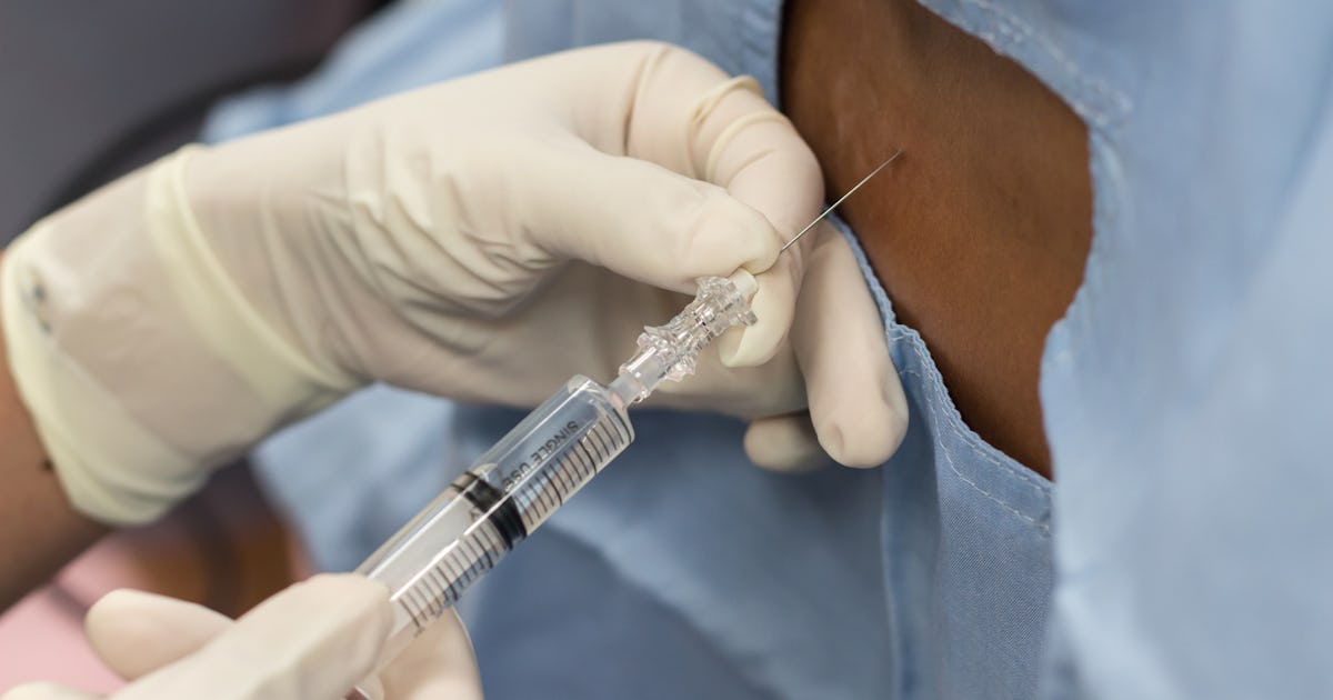
How Long Does An Epidural Last After Birth? You Might Want To Wait A While Before Your First Postpartum Walk

My Labor & Delivery Story | Epidural Didn't work ! - YouTube
:max_bytes(150000):strip_icc()/154725434-56a76f4e5f9b58b7d0ea7922.jpg)
5 Things That Can Help When the Epidural Is Not Working

Why do epidurals not work for some people

Epidural Risk - My Birth Story: 'My Epidural Didn't Work' | Women's Health

Our Birth Story | My Epidural Didn't Work! | Feat. Baby Montana & Margot the Dog | Tiny Acorn - YouTube

I Asked for the Epidural, But Didn't Get What I Expected

Why did no one tell me that epidurals don't always work?

Epidural Steroid Injections Won't Solve Your Back Pain — Pain News Network
How I Found Out Halfway Through Labor I Couldn't Get an Epidural
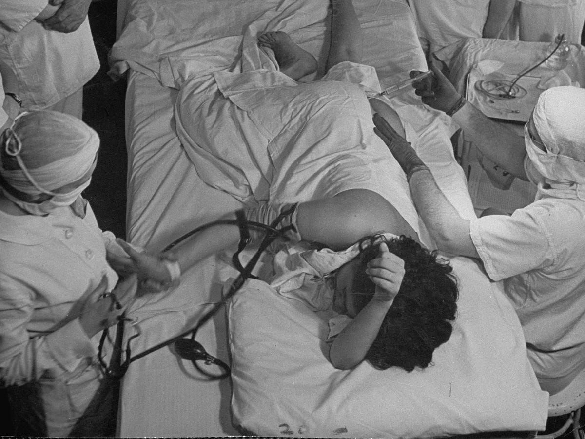
Misogyny and the Epidural: a Primer | by Ellie Slee | Medium
Posting Komentar untuk "epidural didn t work"